2013 Was The Start of Something Big For Neuroscience
 By Michael Q. Bullerdick
Declaring any period of rapid, awe-inspiring achievement a “golden age” is usually best left to scholars—and only then after a little time and some distance. But every now and then it’s easy to make the call at the moment, as was pretty much the case with rocket science, desktop computing and telecommunications. Now, it seems we can make a similar call about the field of neuroscience, where a critical mass of groundbreaking studies is bolstering an international consensus that we are indeed entering a golden age of brain science.
Additional support for such a bold declaration came last year in the form of major European- and U.S-sponsored research grants, established—as President Obama put it so matter-of-factly—“to unlock the mystery of the three pounds of matter between our ears.” In January of 2013, the European Commission established the Human Brain Project, pledging 1 billion euros for a Swiss-based, multi-University effort to construct the most comprehensive functioning computer model of a human brain. In April, President Obama pledged 100 million dollars toward a multi-university study called the BRAIN Initiative (Brain Research through Advancing Innovative Neurotechnologies). The centerpiece of U.S. efforts is the Human Connectome Project (HCP), a first-of-its-kind, ten-year endeavor to catalog and map the human brain—all 100 billion neurons worth, give or take a few. Just as the Human Genome Project famously sequenced human DNA and defined a baseline against which genetic disorders could be diagnosed, HCP will analyze healthy human brains to establish a baseline of “functional connectivity.” From there, disorders such as dyslexia, autism, Alzheimer’s disease and schizophrenia, to name a few, may be better understood and diagnosed.
HCP and, for that matter, our budding “golden age of neuroscience,” are being made possible by a confluence of technologies and a bit of favorable economics. In the last decade or so, supercomputers, like those Amazon relies on to crunch billions of data points in mere seconds, are becoming increasingly available and therefore more affordable. These computational behemoths are necessary to create statistical models of the brain’s billions of neurons, each with a thousand or so connections, as they light up or wink out each second. More importantly, advances in medical resonance imagers (MRIs), like those recently unveiled and in use at Albany Medical Center and the one HCP is utilizing at Massachusetts Medical Center, are making huge gains in navigating the brain’s murky depths. Such sophisticated devices generate images of brain activity and connectivity by tracking blood flow (or water, in the case of more complex diffusion imagers) in the active brains of test subjects: as they sleep, wake, experience emotions or try to suppress them, while they perform routine tasks or even just imagine doing so. That’s a far cry from injecting dyes into dead tissue samples for studying under a microscope.
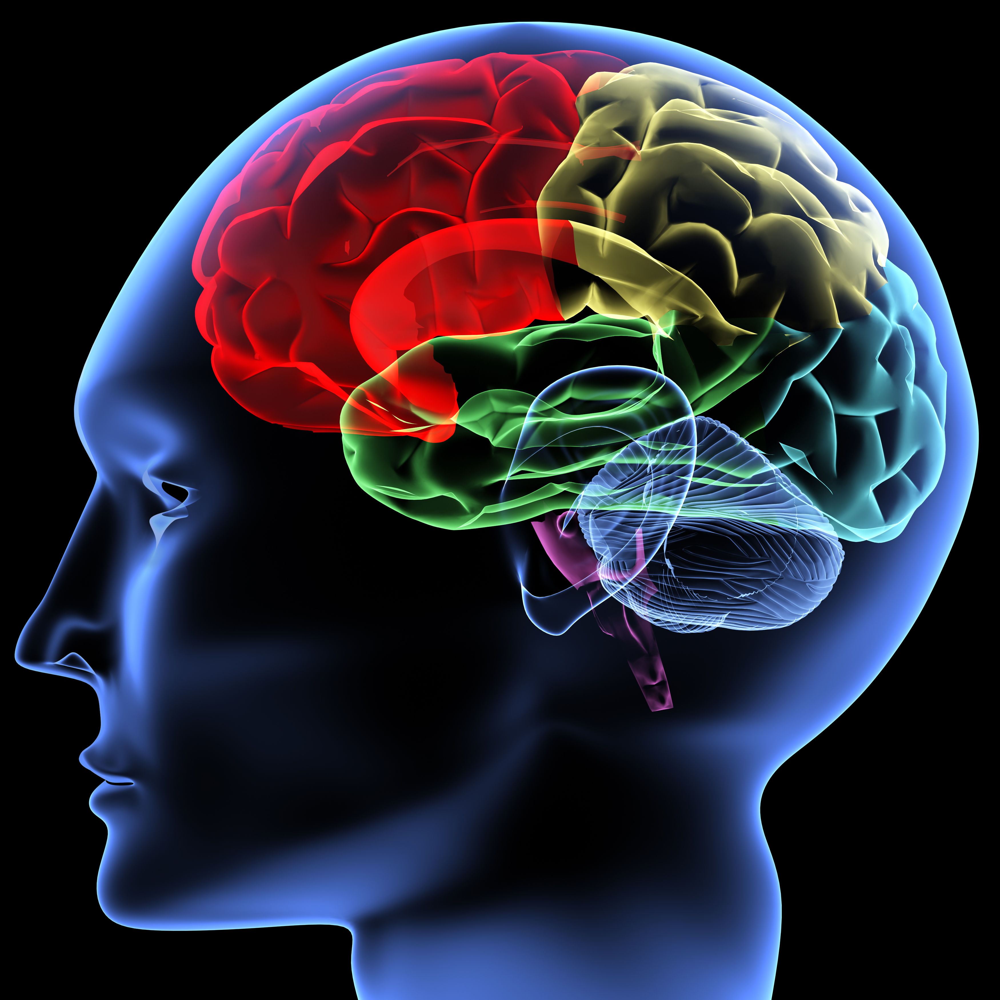
As a result of recent MRI-based studies, researchers are beginning to draw an early schematic of the brain’s complex wiring—how the sizes and shapes of its various components and its neural connectivity impact an individual’s mental makeup and perhaps influence personality.
Last year alone saw the publication of several groundbreaking studies that caused a stir in both academic journals and worldwide media. According to Google analytics, the most widely searched of these stories confirmed what had long been suspected: that the brains of adult males and females differ. In fact, the pioneering study from the University of Pennsylvania, which was published in the Proceedings of National Academy of Sciences, showed that beginning at adolescence, when secondary sex characteristics first manifest, connections in male brains become fixed on the same side of the brain, running from front to back; conversely, those of females run side-to-side between both hemispheres. Although media outlets had a field day linking various gender-based stereotypes to the findings, the study’s researchers were careful to point out that drawing such conclusions was premature. However, they allowed that the findings could be the reason men are better at spacial tasks and activities involving muscle control while women are generally better at language, intuition, and memory-related tasks. Further, the findings may also explain why men are more single minded and women are better at multi-tasking.
Another MRI-based study, conducted by researchers from Tel Aviv University and published in the journal PLOS One, found that patients in a vegetative state might be more aware than has been previously surmised. When such test subjects were spoken to and asked to imagine someone they knew or were shown pictures of friends versus those of strangers, MRI results indicated emotional processing activity for the former but not the latter. The takeaway from this and similar studies is that consciousness may exist as a matter of degrees along a sliding scale and not as a static state, either on or off.
A University of British Columbia study published in Nature Neuroscience found that a little understood region of the brain, the lateral habenula, already known to play a part in regulating depression, also plays a key role in helping us to make decisions. The findings have important implications for the study of how we make choices and how depression may be an influencer.
A study from the University of New Mexico and published in the Proceedings of the National Academy of Sciences found that felons who did poorly on tests designed to measure impulse control demonstrated lower levels of activity in the brain region known to regulate it, the anterior cingulate cortex (ACC). The study also tracked the subjects for several years after their release and noted, perhaps not surprisingly, that those found to have comparatively slow ACCs were four times more likely to be incarcerated again.
Another headline-grabbing study, which utilized subjects suffering from cortical blindness, demonstrated that it is possible to “sense” being watched even when we do not “see” the watcher. MRI readings of cortically blind test subjects showed that the amygdala, the part of the brain that evaluates threat and initiates our fight-or-flight response, activated during periods when they were being stared at by volunteers as opposed to when the volunteers looked away. Since the subjects were blind, the brain activity in both instances should have been unchanged. But the study, from the University of Geneva and published in the Journal of Neuroscience, concluded that brain processing—and therefore a type of perception—can occur even when the visual cortex fails.
These amazing study highlights, all from 2013, represent a tiny speck in the mountain of data that scientists say is fast outpacing the ability of even the most ambitious and well-funded research institutions to review and interpret. The hope is that when they get around to it, and the Human Brain Project and Human Connectome Projects are complete, we’ll be light years closer to understanding the causal relationship between the state of a person’s brain and his actions as a result. The data we already possess suggests a not-so-distant future where diagnosing the extent of brain injuries and complex disorders—currently a lengthy and imperfect process—could be as quick and simple as undergoing a routine x-ray. When and if that day arrives, we’ll no doubt look upon this decade as the golden age of neuroscience.